Extracellular vesicles (EVs) are natural lipid bilayer vesicles, produced by many different cell types and have a plethora of functions and roles in cell-to-cell communication, still under discovery up to now. Our group has deeply studied their role in cancer nanomedicine and immunotherapy, as well as the possibility to engineer these natural vesicles in effective nano-sized carriers for drug or gene delivery and to transport nanoparticles [1-3].
Our idea is to use the lipid bilayers and proteins obtained from EVs as a biomimetic coating to construct our Trojananohorses having intrinsic biomimetic and homing capabilities of natural EVs. The research team has proceeded to the extraction using differential ultracentrifugation and full characterization of the EVs derived from different cell lines, in particular from both cancer and healthy cells cultured in vitro.
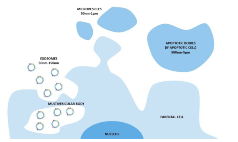
The biogenesis of extracellular vesicles (EVs) and the different pathways according to the current classification. In particular, exosomes consist of vesicles with an endocytic origin, ranging in size from around 50 to 150 nm. They originate as intraluminal vesicles (ILVs) of the multivesicular bodies (MVBs) and become exosomes when secreted in the extracellular milieu. The microvesicles originate from the direct outwards budding and fission of the plasma membrane and range in size from 50 nm to 1 μm. The apoptotic bodies are vesicles resulting from the disassembly of the apoptotic cells, which are generally defined as 500 nm-5 μm in diameter.

The biological role of extracellular vesicles (EVs) was also studied in terms of intrinsic homing capability towards healthy (B lymphocystes) and cancer cells, both Daudi and HL60 cell lines [4]. We also proposed the bioconjugation with the monoclonal antibody in order to have a specific cancer cell targeting.
Related Publications